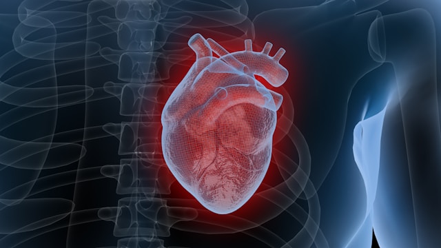
Cardiac catheterization techniques have revolutionized invasive cardiology by offering minimally invasive pathways for both diagnosis and treatment of complex heart conditions. By threading thin, flexible catheters through blood vessels into the heart, clinicians can access chambers and coronary arteries directly. This approach delivers real-time hemodynamic measurements, targeted angioplasty, and precise device placement—all with reduced patient discomfort and faster recovery compared to open-heart surgery. As cardiac catheterization techniques evolve, practitioners leverage advanced imaging, novel catheter materials, and data-driven planning to enhance safety and outcomes. Understanding these innovations is essential for any cardiology team committed to delivering state-of-the-art care.
Evolution of Cardiac Catheterization Techniques
The roots of cardiac catheterization techniques trace back to the 1920s, but it was Nobel Laureates André Cournand and Dickinson Richards who, in the 1950s, standardized hemodynamic assessments in patients. Initially confined to pressure and oxygen saturation monitoring, these techniques rapidly expanded into therapeutic realms by the 1970s with the advent of percutaneous transluminal coronary angioplasty (PTCA). Innovations in polymer science produced catheters with enhanced flexibility and torque control, while refined guidewires enabled access to tortuous vessels. Over subsequent decades, cardiac teams accumulated procedural expertise, integrating fractional flow reserve (FFR) measurements to determine lesion significance and guide revascularization decisions. Today’s operators build on this legacy, combining decades of procedural knowledge with rigorous simulation training to master advanced cardiac catheterization techniques before entering the cath lab.
Advanced Imaging in Cardiac Catheterization Techniques
Fluoroscopy remains the foundational imaging modality in cardiac catheterization techniques, providing real-time X-ray visualization of catheter navigation. However, modern labs now routinely incorporate intravascular ultrasound (IVUS) and optical coherence tomography (OCT) to generate high-resolution, cross-sectional vessel images. These enhancements in visualization allow operators to evaluate plaque composition and stent expansion with unprecedented precision. Additionally, three-dimensional rotational angiography reconstructs complex anatomies, facilitating structural heart procedures such as transcatheter aortic valve replacement (TAVR). Fusion imaging systems—merging pre-procedural CT or MRI data with live fluoroscopy—further optimize guidance, reducing fluoroscopy time and contrast usage. As a result, these cardiac catheterization techniques streamline interventions and minimize radiation exposure for patients and staff.
Next-Gen Materials
The continuous refinement of catheter design is central to advancing cardiac catheterization techniques. Early nylon and polyethylene models have been supplanted by braided, biocompatible polymers that balance flexibility and pushability. Hydrophilic coatings on catheter surfaces reduce friction, facilitating smoother passage through narrow or calcified vessels. Sensor-embedded catheters now deliver instantaneous pressure and flow readings, eliminating the need for multiple device exchanges during a procedure. In therapeutic applications, drug-eluting balloons and bioresorbable scaffolds deliver targeted pharmacotherapy while minimizing long-term foreign body presence in the artery. Steerable sheath systems enable precise access to challenging anatomies, broadening patient eligibility for percutaneous therapies. Together, these material advances ensure that cardiac catheterization techniques remain at the cutting edge of patient-centered innovation.
Precision Patient Care via Cardiac Catheterization Techniques
Implementing cardiac catheterization techniques effectively begins long before the patient enters the lab. Multidisciplinary heart teams review imaging, clinical history, and anatomical considerations to customize procedural plans. During the intervention, FFR and instantaneous wave-free ratio (iFR) guide lesion selection, ensuring revascularization is performed only when hemodynamically warranted. Intraprocedural echocardiography—either transesophageal or intracardiac—confirms device positioning and immediate functional results. Post-procedure, enhanced recovery protocols emphasize early ambulation, rigorous hemodynamic monitoring, and patient education to shorten hospital stays and lower complication rates. Follow-up care utilizes noninvasive CT angiography or stress imaging to detect restenosis or device malfunction promptly. Through these meticulous cardiac catheterization techniques, clinicians elevate patient outcomes and set new standards for safety and efficacy in invasive cardiology.
Innovation in cardiac catheterization techniques continues unabated, driven by a commitment to precision, safety, and improved patient experiences. As imaging modalities become ever more sophisticated and catheter technologies more refined, the scope of percutaneous interventions will expand, offering therapeutic options to patients once deemed inoperable. By staying at the forefront of these advancements, cardiology teams can ensure that each innovation translates into tangible benefits—enhanced diagnostics, reduced procedural risk, and faster, more complete recoveries.